X-ray Crystallography: Revealing the Invisible Architecture of Molecules. Discover How This Technique Transformed Science and Medicine.
- Introduction to X-ray Crystallography
- Historical Milestones and Pioneers
- Fundamental Principles and Physics
- Sample Preparation and Crystal Growth
- Data Collection: X-ray Sources and Detectors
- Solving and Refining Crystal Structures
- Applications in Chemistry and Biology
- Technological Advances and Automation
- Challenges, Limitations, and Error Sources
- Future Directions and Emerging Innovations
- Sources & References
Introduction to X-ray Crystallography
X-ray crystallography is a powerful analytical technique used to determine the atomic and molecular structure of crystalline materials. By directing X-rays at a crystal and analyzing the resulting diffraction patterns, scientists can infer the precise arrangement of atoms within the crystal lattice. This method has been instrumental in advancing our understanding of the structure and function of a wide range of substances, from simple inorganic compounds to complex biological macromolecules such as proteins and nucleic acids.
The origins of X-ray crystallography date back to the early 20th century, following the discovery of X-rays by Wilhelm Röntgen in 1895 and the subsequent demonstration of X-ray diffraction by Max von Laue in 1912. The technique was further developed by William Henry Bragg and William Lawrence Bragg, who formulated Bragg’s Law, providing the theoretical foundation for interpreting X-ray diffraction data. Their pioneering work earned them the Nobel Prize in Physics in 1915 and established X-ray crystallography as a cornerstone of structural science.
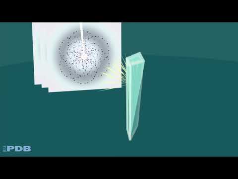
The process of X-ray crystallography involves several key steps. First, a high-quality crystal of the substance under investigation must be obtained. The crystal is then exposed to a focused beam of X-rays, which interact with the electrons in the crystal and are scattered in specific directions. The resulting diffraction pattern is recorded, typically using a detector. By applying mathematical techniques such as Fourier transforms, researchers can reconstruct a three-dimensional electron density map of the crystal, revealing the positions of individual atoms.
X-ray crystallography has had a profound impact on numerous scientific disciplines. In chemistry, it has enabled the elucidation of complex molecular structures, facilitating the design of new materials and pharmaceuticals. In biology, it has been crucial for understanding the architecture of proteins, enzymes, and nucleic acids, including the landmark determination of the double-helix structure of DNA. The technique is widely used in academic research, as well as in industrial and pharmaceutical laboratories around the world.
Several organizations play a central role in the advancement and application of X-ray crystallography. The International Union of Crystallography (IUCr) is a leading authority that promotes international cooperation in crystallography and supports the dissemination of research and standards in the field. Facilities such as synchrotron radiation sources, managed by organizations like the European Synchrotron Radiation Facility (ESRF), provide researchers with access to high-intensity X-ray beams, enabling the study of increasingly complex and challenging samples.
Historical Milestones and Pioneers
X-ray crystallography has played a transformative role in the advancement of structural science since its inception in the early 20th century. The technique’s origins can be traced to 1912, when German physicist Max von Laue demonstrated that crystals could diffract X-rays, providing the first experimental evidence that X-rays are electromagnetic waves and that crystals have a regular, repeating structure. This breakthrough earned von Laue the Nobel Prize in Physics in 1914 and laid the foundation for the field.
Building on von Laue’s discovery, British father-and-son team William Henry Bragg and William Lawrence Bragg developed the mathematical framework to interpret X-ray diffraction patterns. Their formulation, known as Bragg’s Law, enabled scientists to deduce the atomic arrangement within crystals. For this pioneering work, the Braggs were jointly awarded the Nobel Prize in Physics in 1915, making Lawrence Bragg, at age 25, the youngest-ever Nobel laureate in science. The Nobel Prize organization recognizes these achievements as foundational to modern crystallography.
Throughout the 20th century, X-ray crystallography became an indispensable tool for chemists, physicists, and biologists. In 1953, the technique reached a historic milestone when Rosalind Franklin’s X-ray diffraction images of DNA, combined with the model-building efforts of James Watson and Francis Crick, led to the elucidation of the double helix structure of DNA. This discovery revolutionized molecular biology and genetics, and the Research Collaboratory for Structural Bioinformatics (RCSB) Protein Data Bank continues to archive and disseminate structural data derived from X-ray crystallography.
Other notable milestones include the determination of the first protein structure, myoglobin, by John Kendrew and colleagues in 1958, and the subsequent structure of hemoglobin by Max Perutz. These achievements, recognized by the International Union of Crystallography (IUCr), demonstrated the power of X-ray crystallography to reveal the intricate architecture of biological macromolecules.
Today, X-ray crystallography remains a cornerstone of structural science, with ongoing innovations in instrumentation, data analysis, and automation. The technique’s legacy is reflected in the thousands of structures deposited annually in global databases and its continued impact on fields ranging from drug discovery to materials science.
Fundamental Principles and Physics
X-ray crystallography is a powerful analytical technique that reveals the atomic and molecular structure of crystalline materials. The fundamental principle underlying this method is the interaction between X-rays and the periodic lattice of a crystal. When a beam of X-rays, which are electromagnetic waves with wavelengths on the order of 0.01–10 nanometers, is directed at a crystal, the atoms within the crystal cause the X-rays to scatter in specific directions. This scattering is governed by the constructive and destructive interference of the X-ray waves, a phenomenon described by Bragg’s Law. Bragg’s Law, formulated by Sir William Henry Bragg and his son Sir William Lawrence Bragg, states that constructive interference occurs when the path difference between X-rays reflected from successive crystal planes equals an integer multiple of the X-ray wavelength.
Mathematically, Bragg’s Law is expressed as nλ = 2d sinθ, where n is an integer (the order of reflection), λ is the wavelength of the incident X-rays, d is the distance between crystal planes, and θ is the angle of incidence. By measuring the angles and intensities of the diffracted beams, researchers can reconstruct a three-dimensional electron density map of the crystal. This map allows for the determination of the positions of atoms within the unit cell, the smallest repeating unit in the crystal lattice.
The physics of X-ray crystallography relies on the fact that X-rays have wavelengths comparable to interatomic distances, making them ideal for probing crystal structures. When X-rays encounter the electron clouds of atoms, they are elastically scattered, producing a diffraction pattern unique to the arrangement of atoms in the crystal. The resulting pattern is recorded, typically using a detector such as a charge-coupled device (CCD) or a photographic film. The analysis of these patterns requires sophisticated mathematical techniques, including Fourier transforms, to convert the observed diffraction data into a real-space image of the electron density.
X-ray crystallography has been instrumental in advancing fields such as chemistry, biology, and materials science. It has enabled the elucidation of complex biomolecular structures, including proteins and nucleic acids, and has been central to numerous Nobel Prize-winning discoveries. The technique is standardized and supported by major scientific organizations, including the International Union of Crystallography, which promotes the development and application of crystallographic methods worldwide. Additionally, facilities such as synchrotron light sources, managed by organizations like European Synchrotron Radiation Facility, provide high-intensity X-ray beams essential for modern crystallographic studies.
Sample Preparation and Crystal Growth
Sample preparation and crystal growth are foundational steps in X-ray crystallography, directly influencing the quality and interpretability of diffraction data. The process begins with the purification of the target molecule—be it a small organic compound, inorganic material, or a macromolecule such as a protein or nucleic acid. High purity is essential, as contaminants can hinder crystal formation or introduce disorder, complicating structural analysis. For proteins, this often involves recombinant expression systems, followed by chromatographic purification to achieve homogeneity.
Once purified, the sample must be crystallized. Crystal growth is a delicate and often rate-limiting step, particularly for biological macromolecules. The goal is to produce single crystals of sufficient size (typically 0.1–0.5 mm in each dimension) and quality, with minimal defects. Crystallization methods vary depending on the sample type. For small molecules, slow evaporation or cooling of a saturated solution is common. In contrast, proteins and nucleic acids are typically crystallized using vapor diffusion (hanging or sitting drop), microbatch, or dialysis techniques. These methods manipulate parameters such as pH, temperature, precipitant concentration, and additives to promote nucleation and subsequent crystal growth.
Optimization of crystallization conditions is often empirical, requiring systematic screening of hundreds or thousands of conditions. Robotic systems and high-throughput screening platforms have become invaluable, enabling parallel testing of diverse conditions with minimal sample consumption. Organizations such as the European Molecular Biology Laboratory and RCSB Protein Data Bank provide resources, protocols, and databases to support crystallographers in this endeavor.
Once crystals are obtained, they must be harvested and mounted for X-ray exposure. This step may involve cryoprotection—soaking crystals in solutions containing cryoprotectants (e.g., glycerol or ethylene glycol)—to prevent ice formation during flash-cooling in liquid nitrogen. Proper handling is critical to preserve crystal integrity and minimize radiation damage during data collection. The International Union of Crystallography, a leading authority in the field, offers guidelines and best practices for sample preparation, crystal handling, and data collection.
In summary, meticulous sample preparation and crystal growth are prerequisites for successful X-ray crystallography. Advances in automation, screening technologies, and community resources continue to improve the efficiency and success rate of this crucial phase, enabling the determination of increasingly complex structures.
Data Collection: X-ray Sources and Detectors
Data collection is a critical phase in X-ray crystallography, as the quality and accuracy of the resulting structural information depend heavily on the characteristics of the X-ray sources and detectors employed. The process begins with the generation of X-rays, which are directed at a crystallized sample. The interaction between the X-rays and the crystal lattice produces a diffraction pattern, which is then captured by specialized detectors for subsequent analysis.
Historically, X-ray tubes were the primary sources of X-rays in crystallography. These devices generate X-rays by bombarding a metal target, typically copper or molybdenum, with high-energy electrons. While X-ray tubes remain widely used in laboratory settings due to their accessibility and ease of operation, they are limited in terms of intensity and brilliance. To overcome these limitations, synchrotron radiation facilities have become increasingly important. Synchrotrons are large-scale research infrastructures that accelerate electrons to nearly the speed of light, producing extremely bright and tunable X-ray beams. The high brilliance and collimation of synchrotron X-rays enable the study of very small crystals and facilitate time-resolved experiments. Leading synchrotron facilities include the European Synchrotron Radiation Facility, Advanced Photon Source, and Diamond Light Source, each providing access to state-of-the-art beamlines for crystallographic research.
The choice of detector is equally crucial for accurate data collection. Early crystallography experiments relied on photographic film, but modern laboratories now use electronic detectors that offer higher sensitivity, faster readout, and greater dynamic range. Charge-coupled device (CCD) detectors were once the standard, but have largely been supplanted by pixel array detectors (PADs), such as those based on hybrid photon counting technology. These detectors, exemplified by devices from DECTRIS, provide rapid data acquisition, low noise, and high spatial resolution, making them ideal for both routine and advanced crystallographic studies.
The integration of advanced X-ray sources and detectors has revolutionized data collection in X-ray crystallography. High-brilliance synchrotron sources, combined with fast, sensitive detectors, allow researchers to collect complete datasets from tiny or weakly diffracting crystals, and to perform experiments that probe dynamic structural changes. These technological advancements continue to expand the frontiers of structural biology, materials science, and chemistry.
Solving and Refining Crystal Structures
Solving and refining crystal structures are central steps in the process of X-ray crystallography, a technique that enables the determination of the three-dimensional arrangement of atoms within a crystalline material. Once a suitable crystal has been obtained and exposed to X-ray radiation, the resulting diffraction pattern is collected. The first major challenge is to solve the so-called “phase problem,” as only the intensities of the diffracted beams are measured, not their phases. Several methods exist to address this, including direct methods, Patterson methods, and molecular replacement, each suited to different types of crystals and data quality.
After initial phase determination, an electron density map is generated, which provides a three-dimensional representation of where electrons are most likely to be found within the unit cell. This map serves as the basis for building an initial atomic model of the molecule or material under investigation. The process of model building is iterative and often involves both automated algorithms and manual intervention, especially in complex biological macromolecules.
Refinement is the subsequent step, where the preliminary model is adjusted to best fit the observed diffraction data. This involves optimizing parameters such as atomic positions, thermal vibrations (B-factors), and occupancies. The goal is to minimize the difference between the observed and calculated structure factors, typically using least-squares or maximum likelihood methods. Modern refinement software incorporates restraints and constraints to ensure chemically reasonable geometry, and validation tools are used to assess the quality of the final model.
Throughout the process, crystallographers rely on specialized software and databases. The International Union of Crystallography (IUCr) plays a pivotal role in setting standards for data collection, structure validation, and publication. The Research Collaboratory for Structural Bioinformatics (RCSB), which manages the Protein Data Bank (PDB), is a key resource for depositing and accessing macromolecular structures. For small molecules, the Cambridge Crystallographic Data Centre (CCDC) maintains the Cambridge Structural Database (CSD), a comprehensive repository of crystal structures.
The accuracy and reliability of a crystal structure depend on the quality of the diffraction data, the resolution achieved, and the rigor of the refinement process. Advances in computational methods, detector technology, and synchrotron radiation sources have significantly enhanced the precision and throughput of structure determination. As a result, X-ray crystallography remains an indispensable tool in chemistry, materials science, and structural biology for elucidating molecular architecture and guiding functional insights.
Applications in Chemistry and Biology
X-ray crystallography is a cornerstone analytical technique in both chemistry and biology, enabling the detailed visualization of molecular and atomic structures. Its primary application lies in determining the three-dimensional arrangement of atoms within crystalline materials, which has profound implications for understanding chemical bonding, molecular geometry, and biological function.
In chemistry, X-ray crystallography is indispensable for elucidating the structures of small organic and inorganic molecules. By analyzing the diffraction patterns produced when X-rays interact with a crystal, chemists can precisely determine bond lengths, bond angles, and the overall molecular conformation. This information is critical for verifying the outcomes of synthetic reactions, characterizing new compounds, and studying reaction mechanisms. The technique has also been pivotal in the development of materials science, aiding in the design of novel catalysts, polymers, and nanomaterials with tailored properties.
In the realm of biology, X-ray crystallography has revolutionized our understanding of macromolecular structures, particularly proteins and nucleic acids. The technique was instrumental in the discovery of the double helix structure of DNA, a milestone that transformed molecular biology. Today, it remains the gold standard for high-resolution structural determination of proteins, enzymes, and large biological complexes. By revealing the precise arrangement of amino acids and active sites, X-ray crystallography provides insights into protein function, mechanisms of enzyme catalysis, and the molecular basis of diseases.
One of the most impactful applications in biology is structure-based drug design. Pharmaceutical researchers use X-ray crystallography to visualize how potential drug molecules interact with their biological targets at the atomic level. This structural information guides the optimization of drug candidates, improving efficacy and reducing side effects. Many life-saving medications, including antiviral drugs and cancer therapeutics, have been developed with the aid of crystallographic data.
The technique is supported and advanced by major scientific organizations and facilities worldwide. For example, International Union of Crystallography (IUCr) promotes the development and application of crystallographic methods, while large-scale synchrotron facilities such as those operated by European Synchrotron Radiation Facility and Argonne National Laboratory provide high-intensity X-ray sources essential for studying challenging biological and chemical samples. These organizations play a crucial role in training researchers, developing new methodologies, and maintaining databases of crystallographic structures.
In summary, X-ray crystallography is a foundational tool in chemistry and biology, enabling discoveries that drive innovation in science, medicine, and technology.
Technological Advances and Automation
X-ray crystallography has undergone significant transformation in recent decades, driven by technological advances and the integration of automation. These developments have dramatically increased the speed, accuracy, and accessibility of structural determination for a wide range of biological and chemical molecules.
One of the most impactful advances is the evolution of X-ray sources. The introduction of synchrotron radiation facilities has provided researchers with highly intense and tunable X-ray beams, enabling the collection of high-resolution diffraction data from even the smallest or most weakly diffracting crystals. Synchrotrons, such as those operated by European Synchrotron Radiation Facility and Advanced Photon Source, have become essential resources for the global crystallography community. More recently, X-ray free-electron lasers (XFELs) have enabled the study of dynamic processes and radiation-sensitive samples by delivering ultrafast, extremely bright pulses, as seen at facilities like SLAC National Accelerator Laboratory.
Automation has revolutionized nearly every stage of the crystallographic workflow. Robotic systems now handle high-throughput crystallization screening, crystal mounting, and data collection, minimizing human error and increasing reproducibility. Automated sample changers and goniometers, integrated with advanced software, allow for remote and unattended data acquisition, which is particularly valuable at large-scale facilities. The development of sophisticated data processing pipelines, such as those supported by International Union of Crystallography and implemented in software like CCP4 and PHENIX, has streamlined the conversion of raw diffraction images into interpretable electron density maps and atomic models.
Recent advances in detector technology, such as pixel array detectors, have further enhanced data quality and collection speed. These detectors offer high sensitivity, rapid readout, and low noise, making them ideal for both synchrotron and laboratory-based X-ray sources. Additionally, machine learning and artificial intelligence are increasingly being applied to automate crystal identification, optimize data collection strategies, and improve model building and validation.
Collectively, these technological and automation advances have made X-ray crystallography more efficient and accessible, enabling researchers to tackle increasingly complex biological questions and accelerating the pace of discovery in structural biology, materials science, and drug development.
Challenges, Limitations, and Error Sources
X-ray crystallography has been a cornerstone technique in structural biology, chemistry, and materials science, yet it faces several intrinsic challenges, limitations, and sources of error that can impact the accuracy and reliability of its results. One of the primary challenges is the requirement for high-quality crystals. Many biologically relevant molecules, such as membrane proteins and large macromolecular complexes, are notoriously difficult to crystallize, which restricts the applicability of the method. The process of crystallization itself can introduce artifacts, as the conditions necessary for crystal formation may induce non-physiological conformations or packing interactions that do not reflect the native state of the molecule.
Another significant limitation is the phase problem. While X-ray diffraction provides information about the amplitude of scattered waves, it does not directly yield phase information, which is essential for constructing accurate electron density maps. Various methods, such as multiple isomorphous replacement and anomalous dispersion, have been developed to address this, but they add complexity and potential for error to the process. Additionally, the resolution of the resulting structure is limited by the quality of the crystal and the data collected. Poorly ordered crystals or those with high mosaicity can lead to low-resolution data, making it difficult to model atomic positions with confidence.
Radiation damage is another source of error, particularly for sensitive biological samples. Prolonged exposure to X-rays can cause chemical changes or breakage of bonds within the crystal, leading to artifacts in the resulting structure. Cryo-cooling techniques are commonly used to mitigate this, but they do not eliminate the problem entirely. Furthermore, model bias can occur during the interpretation of electron density maps, especially when prior knowledge or expectations influence the fitting of atomic models.
Errors can also arise from data processing and refinement. Inaccurate scaling, incorrect space group assignment, or improper handling of symmetry can introduce systematic errors. The validation of structures is therefore critical, and organizations such as the Worldwide Protein Data Bank (wwPDB) play a key role in setting standards for data deposition, validation, and dissemination. The International Union of Crystallography (IUCr) also provides guidelines and resources to promote best practices in crystallographic research.
In summary, while X-ray crystallography remains a powerful and widely used technique, its effectiveness is constrained by challenges in crystallization, phase determination, radiation damage, and data interpretation. Ongoing advances in instrumentation, computational methods, and community standards continue to address these limitations, but careful experimental design and critical evaluation of results remain essential for reliable structural determination.
Future Directions and Emerging Innovations
X-ray crystallography, a cornerstone of structural biology and materials science, continues to evolve with technological advancements and interdisciplinary integration. The future of this technique is shaped by innovations aimed at overcoming traditional limitations, such as the need for large, well-ordered crystals and the challenges of studying dynamic or complex biological systems.
One significant direction is the development of serial femtosecond crystallography (SFX) using X-ray free-electron lasers (XFELs). SFX enables the collection of diffraction data from micro- or nanocrystals using ultrafast, intense X-ray pulses, capturing structural information before radiation damage occurs. This approach is particularly valuable for studying proteins that are difficult to crystallize in large forms or are sensitive to radiation. Facilities like the European XFEL and SLAC National Accelerator Laboratory are at the forefront of this innovation, providing researchers with access to cutting-edge XFEL sources.
Another emerging trend is the integration of cryo-electron microscopy (cryo-EM) and X-ray crystallography. By combining high-resolution crystallographic data with cryo-EM maps, scientists can build more complete models of large macromolecular complexes and membrane proteins. This hybrid approach leverages the strengths of both techniques, expanding the range of biological questions that can be addressed.
Advances in computational methods are also transforming X-ray crystallography. Machine learning algorithms and artificial intelligence are being applied to automate crystal identification, optimize data collection strategies, and improve phase determination. These tools accelerate the structure determination process and enhance the accuracy of resulting models. Organizations such as International Union of Crystallography are actively supporting the development and dissemination of these computational resources.
Miniaturization and automation are making crystallography more accessible. Microfluidic devices and robotic systems now enable high-throughput crystallization screening and data collection, reducing sample consumption and increasing efficiency. This is particularly beneficial for drug discovery, where rapid screening of protein-ligand complexes is essential.
Looking ahead, the integration of X-ray crystallography with complementary techniques—such as neutron diffraction, spectroscopy, and in situ studies—promises to provide deeper insights into dynamic processes and functional mechanisms at the atomic level. As synchrotron and XFEL facilities continue to expand their capabilities, X-ray crystallography is poised to remain a vital tool in structural science, driving discoveries in biology, chemistry, and materials research.
Sources & References
- International Union of Crystallography
- Nobel Prize
- Research Collaboratory for Structural Bioinformatics (RCSB) Protein Data Bank
- European Molecular Biology Laboratory
- European Synchrotron Radiation Facility
- Advanced Photon Source
- DECTRIS
- Cambridge Crystallographic Data Centre
- Worldwide Protein Data Bank
- European XFEL